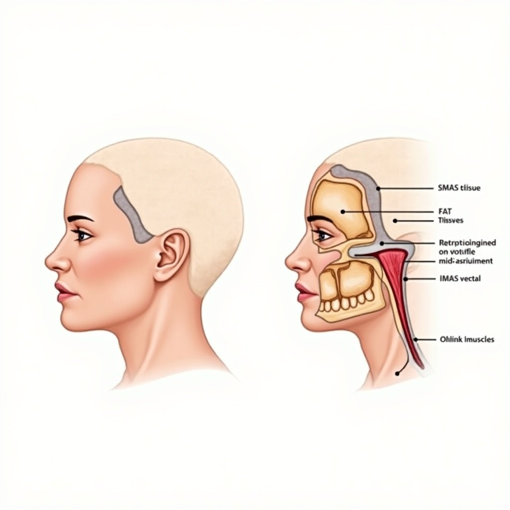
Educational Illustration of Deep Plane Facial Lift Technique with SMAS and Mid-Face Repositioning
A
Generated by FLUX.1-dev
G
Image Size: 1024 x 1024
Flux AI Model: FLUX.1-dev
Generator: Square
Flux Prompt
AI Prompt
More Flux Images About Deep plane facial lift technique illustration
Educational Illustration of Deep Plane Facial Lift Technique with SMAS and Mid-Face Repositioning and Related Flux Artwork

A


D


T


