Vascular Cross-Section

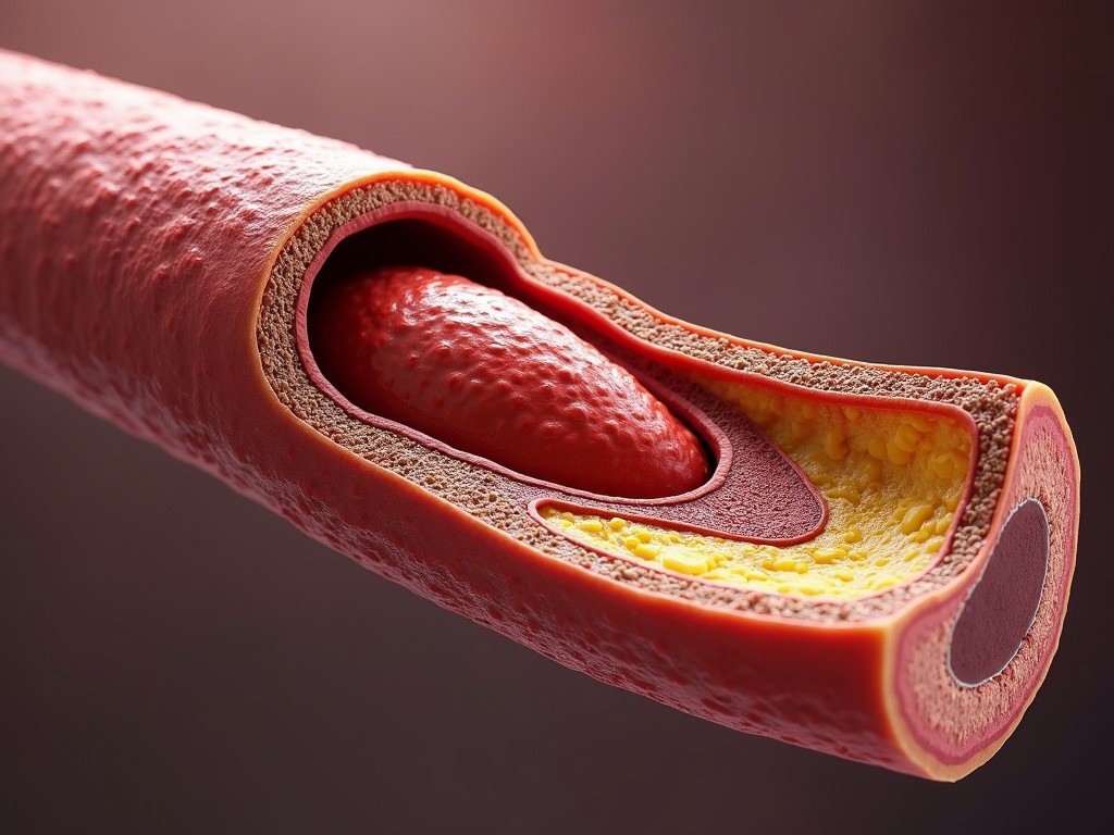
This image presents a highly detailed cross-sectional view of an artery showing the layers and structure of the blood vessel. The artery’s wall is cut away to reveal the buildup of plaque within the vessel, a key feature in understanding cardiovascular diseases like atherosclerosis. The image is designed to illustrate the narrowing of the artery due to lipid deposits, which could lead to restricted blood flow.
m

Generated by FLUX.1-pro

G

Flux Prompt

AI Prompt

More Flux Images About Cross-section of an artery

Vascular Cross-Section and Related Flux Artwork

This image presents a highly detailed cross-sectional view of an artery showing the layers and structure of the blood vessel. The artery’s wall is cut away to reveal the buildup of plaque within the vessel, a key feature in understanding cardiovascular diseases like atherosclerosis. The image is designed to illustrate the narrowing of the artery due to lipid deposits, which could lead to restricted blood flow. This visual aid is beneficial for educational purposes, showcasing arterial health. It's ideal for medical professionals and students to understand the implications of plaque formation.
m

FLUX.1-pro AI

Detailed Cross-Section of an Artery Illustrating Plaque Buildup and Cardiovascular Health

This image presents a highly detailed cross-sectional view of an aneurysm artery. The artery's wall is cut away to reveal the buildup of plaque within the vessel. Understanding this buildup is crucial for studying cardiovascular diseases like atherosclerosis. The layers of the artery are intricately displayed, showcasing its structural composition. Such depictions are essential for educational purposes in medicine and biology.
m

FLUX pro AI

Detailed Cross-Section of an Aneurysm Artery Highlighting Plaque Buildup

The image depicts a cross-section of an artery, filled with blood cells and covered with a layer that resembles atherosclerosis. The inner surface appears smooth, with irregularly shaped particles suggesting a depiction of arterial plaque. The labels, possibly indicating anatomical features, are artistically styled bubbles pointing to different areas.
2 images
m

FLUX.1-pro Image

Internal Flow

This image provides a close-up view inside a human artery. It depicts a cross-section that reveals the layers of the arterial wall, showing a build-up of cholesterol or plaque deposits that threaten to impede blood flow. The textures and colors capture the intricate biological structures in vivid detail, emphasizing the health risks associated with arterial blockages.
m

Flux Image Generator

Inside the Artery: A Close-up View

The image is a detailed and colorful 3D illustration showcasing a cross-section of an artery with visible cholesterol build-up. The artery walls are depicted in shades of red, yellow, and orange, illustrating layers of the tissue and fat deposits. The focus is on the dark, round shapes representing blood cells, emphasizing the narrowing of the artery due to plaque accumulation.
m

FLUX pro Image Generator

Artery Cross-Section - Cholesterol Build-Up

This image presents an intricate view of a human artery cross-section, highlighting the layers and accumulation of cholesterol within. The interior is filled with a yellowish substance, representing plaque buildup, which contrasts against the reddish-brown, textured exterior of the arterial wall. The close-up perspective reveals the biological complexity and detail of the vascular structure, potentially serving as a medical or educational illustration.
m

FLUX pro Image Generator

Intricate Arterial Cross-Section

This image is a detailed cross-section of a human artery, illustrating plaque buildup, a common cause of cardiovascular disease. The artery's inner layers can be seen, with a visible accumulation of yellowish plaque material on the artery walls, reducing the passage for blood flow. This visualization highlights the importance of monitoring and maintaining cardiovascular health.
m

Flux Image Generator

Arterial Insight

The image depicts a detailed cross-section of a clogged artery, showcasing various layers, including cholesterol build-up. The vibrantly colored and textured artery wall contrasts with the yellowish plaque deposits, creating a visually striking representation. The image effectively illustrates the concept of cardiovascular health challenges.
m

Flux AI

Inside the Artery

The illustration depicts the intricate design of an intravascular stent. It features a two-layer structure: the outer layer acts as an anchor, while the inner layer contains the electrodes and sensors. The drawing clearly distinguishes between the two layers and their connection points. Fine details highlight the engineering behind the stent's functionality. This visual serves an educational purpose in the field of biomedical engineering.
K

Flux AI

Detailed Illustration of Intravascular Stent Design Featuring Two-Layer Structure with Electrodes and Sensors

The image presents a detailed cross-sectional view of a human artery, illustrating the buildup of cholesterol plaque along the inner walls. Blood cells can be seen traveling through the arterial passage, conveying the process of atherosclerosis, where plaque accumulation narrows the artery and restricts blood flow. The visual effectively captures the medical concept in a vivid and clear manner.
m

FLUX pro AI

Journey of Blood Cells

Illustration of a cross-section of a cell with colorful organelles
2 images
I

FLUX.1-pro AI

Vibrant Cellular Cross-Section

The image is a schematic diagram showing a layout of three interconnected units, each labeled with components like 'MULTIPLEXER' and 'ASYNC'. Connecting lines indicate data flow or communication pathways among these units. Labels such as 'Demodulator' and 'CP A/P M4' suggest a technical, engineering context, possibly related to signal processing or telecommunications.
N

Flux Image Generator

Network Diagram Illustration