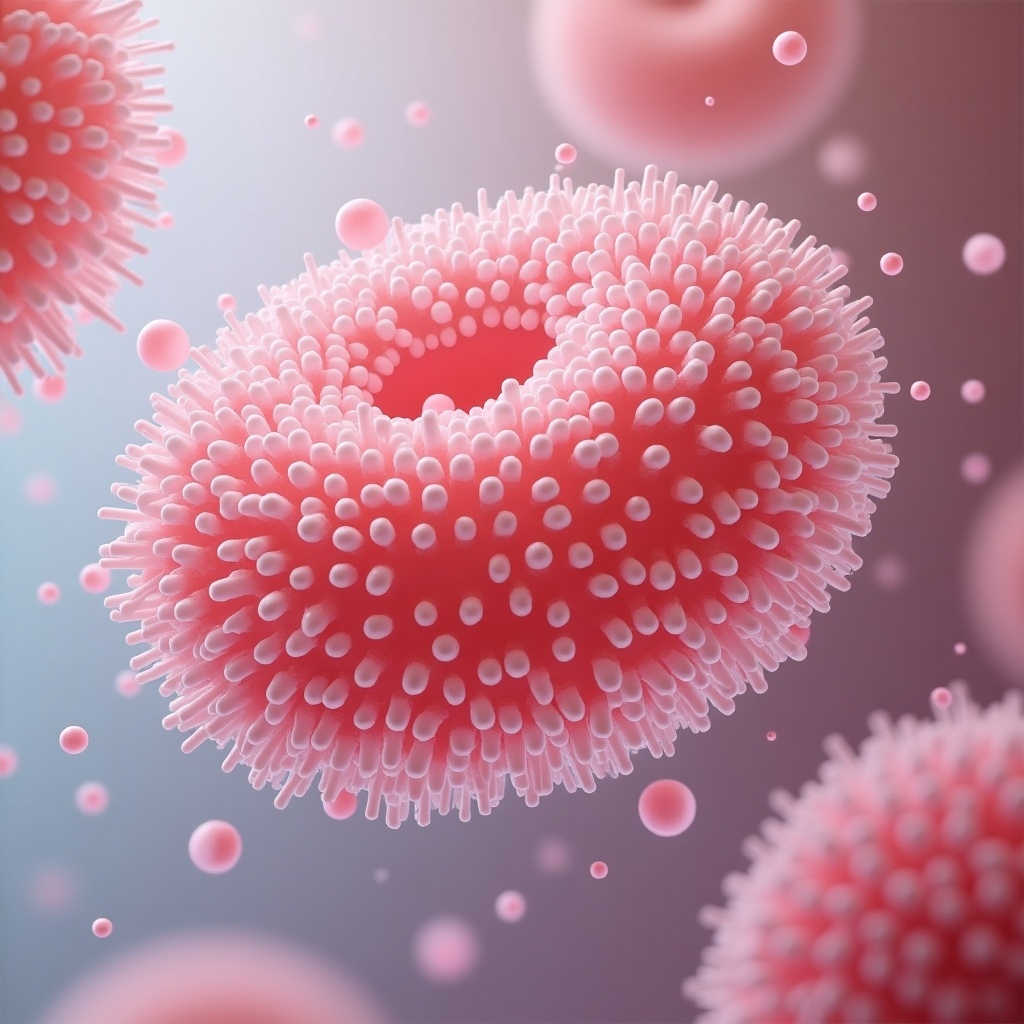
Enhanced Blood Compatibility of Polyurethane Through Sulfate Alginate Modification - Microscopic View
Flux Prompt

AI Prompt

More Flux Images About Microscopic representation of modified polyurethane with sulfate alginate

Enhanced Blood Compatibility of Polyurethane Through Sulfate Alginate Modification - Microscopic View and Related Flux Artwork

a 3D rendered close-up view of a virus particle with red spikes and a blue background
I

FLUX.1-dev Image

Microscopic Intricacies

This image depicts pancreatic beta cells, characterized by their pink, fluffy appearance, which are being shielded from cytokine-induced inflammation. The cells are surrounded by smaller structures that represent cytokines or inflammatory markers. Glowing orange highlights are illustrated within the cells to signify activity or protection by HDAC inhibitors. The dark blue background enhances the contrast, emphasizing the primary subjects. This visual serves as an educational representation of the cellular interactions relevant to diabetes treatment and research.
A

FLUX.1-dev AI

Protective Role of HDAC Inhibitors in Pancreatic Beta Cells Against Cytokine Induced Inflammation

The image showcases two round, shiny pearls positioned side by side. Viewed through a magnifying glass, the viewer can see parallel axes highlighted between the pearls. The angle formed by these axes is distinct and prominent. This representation illustrates the concept of inelastic neutron scattering. The background is a soft white, emphasizing the clarity of the pearls and their reflections. The overall composition is designed to be both visually appealing and scientifically instructive.
J

Flux Image Generator

Exploring Inelastic Neutron Scattering: Pearls Through a Magnifying Glass

create a scientific illustration of Deucravacitinib depicting cellular interactions with labeled elements
A

FLUX.1-pro AI

Scientific Illustration of Deucravacitinib Mechanism Depicting Cellular Interactions

The image shows a newborn baby with a rare medical condition called harlequin ichthyosis. The baby's skin appears red and shiny, covered in large, thick, diamond-shaped plates of skin. These scales make the skin look tight and shiny. The baby is swaddled in green medical cloth, and a gloved hand can be seen in the background, indicating a clinical setting. This condition requires immediate medical attention and specialized care.
2 images
S

FLUX pro AI

Newborn Baby with Harlequin Ichthyosis: A Rare Medical Condition

This image depicts a cross-section of a model of a human artery with a significant buildup of plaque. The artery has been cut open to reveal a large, dark obstruction, likely representing a blood clot or severe cholesterol buildup, which is surrounded by yellowish plaque along the arterial walls. The model uses vibrant colors to highlight the different components and is set against a plain white background.
m

Flux AI Image Generator

Obstructed Artery Model

An illustration depicting a step-by-step scientific process using a polymer solution, with diagrams of mixing, fiber production, and an end product, labeled with technical terms.
S

FLUX pro AI

Illustrated Polymer Process Diagram

A close-up view of a tarantula, highlighting its striking black and orange-colored hairs.
5 images
E

FLUX.1-schnell Image

Majestic Tarantula in Focus

The image shows a pair of hands wearing blue medical gloves, holding a dental prosthetic. The prosthetic includes a set of upper teeth and a pink gum-like base. Metal fasteners are visible on the upper part of the prosthetic, suggesting it's designed to be attached to dental implants. The background is dark, highlighting the dental appliance. This image appears to be related to dental health or dentistry.
2 images
G

Flux AI Image

Close-Up of Dental Prosthetic with Metal Fasteners Held by Gloved Hands

This image presents a close-up view of a cellular structure, reminiscent of bacteria or fungi. The organism features a spiky, textured surface, showcasing intricate details that highlight its biological nature. The dominant colors are teal and green, giving it a vibrant appearance against a softly blurred background. This visualization could be used in various scientific contexts, including education and research. Its macro perspective emphasizes the complexity of microscopic life forms, making it suitable for academic purposes.
O

FLUX.1-pro Image

Close-Up Digital Illustration of a Microbial Structure with Spiky Texture

This image illustrates an experiment involving three groups of rats to study liver preservation techniques. Group 1 is subjected to Ringer lactate washing and immediate ex vivo perfusion. Group 2 uses the IGL-1 solution for cold storage for 24 hours followed by perfusion. Group 3 incorporates DHN-5 into the IGL-1 solution before the same cold storage and perfusion steps. Each group consists of eight rats, clearly labeled. The experiment is set in a laboratory environment with a focus on scientific accuracy and educational value. The jars provide a clear view of the rats and their respective treatments.
J

FLUX.1-pro Image

Experimental Study of Liver Preservation Techniques in Rats using IGL-1 and DHN-5 Solutions

This image is a detailed cross-section of a human artery, illustrating plaque buildup, a common cause of cardiovascular disease. The artery's inner layers can be seen, with a visible accumulation of yellowish plaque material on the artery walls, reducing the passage for blood flow. This visualization highlights the importance of monitoring and maintaining cardiovascular health.
m

Flux Image Generator

Arterial Insight